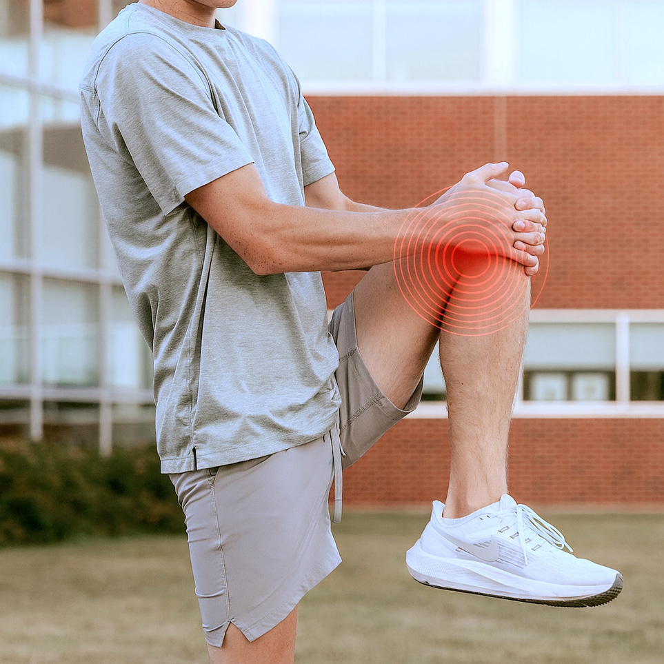
MRI Knee

5 395 kr

MRI Knee

Magnetic Resonance Imaging

Information

Medical comment included

With us, you will always receive a doctor’s opinion from a licensed physician when you order a medical check-up that includes a medical opinion. The doctor’s opinion includes an overall assessment of your results from laboratory analysis. The opinion is individual and may vary depending on the specific tests your medical check-up includes.

What can I expect in the medical report?

The medical opinion will summarize the results of your test results to give you an assessment of your health including any abnormalities outside the reference ranges. In case of abnormalities or need, your doctor may also contact you for a free conversation to explain and talk about your results. Remember that a health check always includes specific test results based on your health and your personal circumstances. Values outside the reference range should therefore always be discussed with a doctor before any decisions regarding possible treatment are made.

What does the doctor look at during a health check?

When assessing a blood test or health check, your doctor will look at several different parameters to evaluate your health and identify any abnormalities. Below are some common aspects your doctor may examine:

Blood counts: Your doctor will check red, white and platelet counts to identify any signs of anaemia, infection or bleeding.

Nutrients and biochemical markers: Includes, for example, analysis and monitoring of blood sugar (glucose) levels, electrolyte levels (e.g. sodium, potassium), liver function tests (e.g. liver enzymes), kidney function tests (e.g. creatinine, urea) and lipid profile (e.g. cholesterol, triglycerides). The results of the tests give your doctor a better idea of your organ function, blood sugar level, cholesterol conditions and other important health aspects.

Inflammatory markers: Your doctor may measure health markers such as C-reactive protein (CRP) or red blood cell decrease rate (SR) to evaluate inflammatory conditions or confirm an active infection.

Hormone levels: Your doctor may monitor your hormone levels to assess hormonal imbalances or diseases, such as thyroid hormones (TSH, T3, T4), sex hormones or other specific hormones such as testosterone levels.

Specific tests: Depending on your goal of your health check or medical history, your doctor may look into specific tests to investigate conditions or diseases, such as diabetes, vitamin deficiency or autoimmune diseases.

In conclusion, the doctor weighs all test results based on the health check to assess your overall health, detect any abnormalities and identify possible underlying medical conditions.

What happens if I have abnormal test results?

If your blood count is abnormal, this will be addressed in your personal medical opinion. Depending on the nature and extent of the abnormality, your doctor may also contact you directly through the test result service for further guidance. Various measures and advice will be provided based on the specific abnormalities observed along with your medical history.

Call included

We always offer a phone consultation with a licensed physician before a referral for an MRI or ultrasound is issued. This allows us to ask the right questions and ensure that you receive the most appropriate and accurate examination for your needs. After the examination, you will receive both the radiologist’s report and a medical opinion, and we remain available to support you throughout the entire process. Follow-up consultations can be booked if desired. Once you have activated the referral, one of our licensed physicians will call you.


Any questions? Call us for advice at 08–14 24 49.

5 395 kr
  • MRI knee – advanced magnetic resonance imaging without referral.
  • Recommended for knee pain, instability, or suspected injury.
  • High-precision imaging of meniscus, cruciate ligaments, cartilage, and joint structures<./li>
  • Fast, painless examination with specialist evaluation within a few days.
Read more

Add-ons

Affiliate Icon

Here it is empty.

Try removing filters or reloading.
Reload
Lågt pris
Lowest price on the market
Ingen strålning
No radiation
Ingen strålning
Referral within 1 business day

MRI Knee – detailed imaging for knee pain and suspected injury

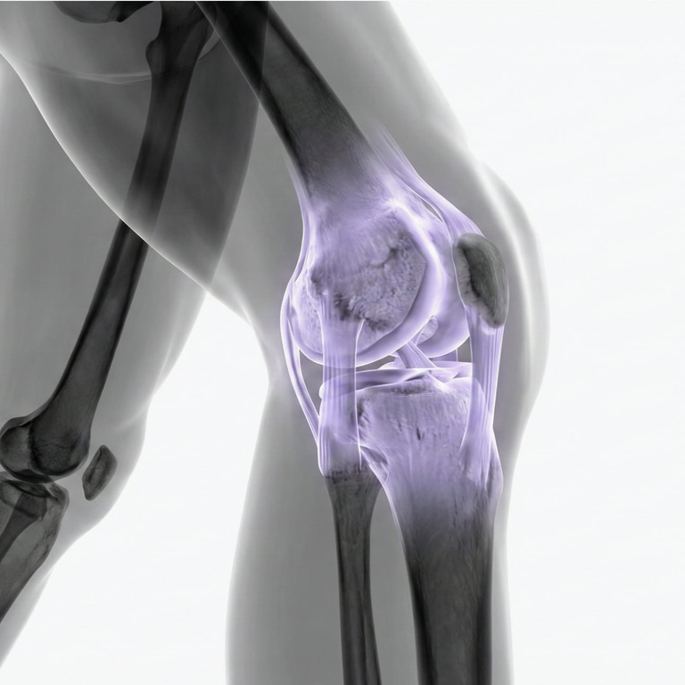
An MRI of the knee provides detailed images of the knee joint and surrounding soft tissues. The examination can assess the menisci, cruciate ligaments, collateral ligaments, cartilage, tendons, bone marrow, and other structures that may be difficult to evaluate with standard X-rays.

Knee MRI may be appropriate for persistent or recurring knee pain, swelling, instability, or after a twisting injury or sports-related trauma where an internal knee injury is suspected. MRI does not use ionising radiation.

The most appropriate imaging method depends on your symptoms and how the problem started. A medical assessment is therefore included to help determine whether a knee MRI is the right examination for your specific symptoms and clinical question.
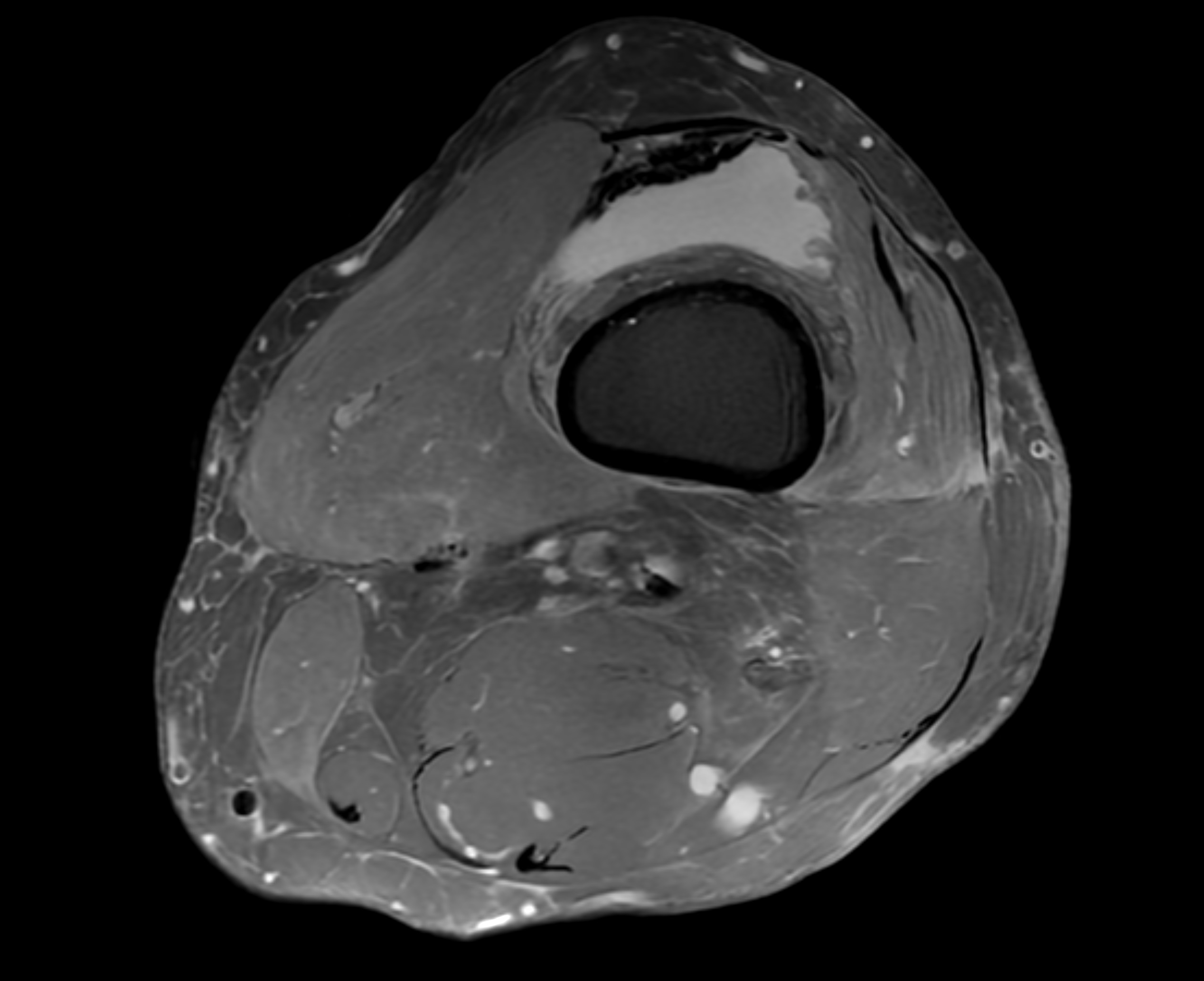
MRI knee – magnetic resonance imaging of the knee joint

Knee pain – when can an MRI be helpful?

Knee pain is common and can have many different causes. Symptoms may arise from the menisci, ligaments, cartilage, tendons, bone, or other structures in and around the knee joint.

An MRI is not necessary for every type of knee pain. Short-term symptoms may be better assessed initially with a clinical examination, rehabilitation, or another imaging method. MRI can be particularly useful when symptoms persist or when there is suspicion of an internal knee injury that cannot be fully evaluated through clinical examination or plain X-ray.

An MRI of the knee may be appropriate for:

  • Persistent or recurring knee pain.
  • Pain after a twisting injury, fall, or sports injury.
  • Swelling or recurrent fluid accumulation in the knee.
  • Instability or a feeling that the knee gives way.
  • Locking or significant mechanical symptoms.
  • Suspected meniscal injury.
  • Suspected ACL, PCL, or other ligament injury.
  • Suspected cartilage or osteochondral injury.
  • Unexplained symptoms where previous examinations have not provided enough information.
  • Follow-up of certain previously diagnosed knee injuries or abnormalities.

What can an MRI of the knee show?

MRI provides detailed images of both soft tissues and the internal structure of bone. This makes it useful for assessing several important parts of the knee joint.

  • The medial and lateral meniscus.
  • The anterior and posterior cruciate ligaments, ACL and PCL.
  • The medial and lateral collateral ligaments, MCL and LCL.
  • Articular cartilage and joint surfaces.
  • The kneecap and structures around the patellofemoral joint.
  • Tendons and other soft tissues around the knee.
  • Bone marrow changes, including bone marrow oedema and stress reactions.
  • Joint fluid, synovitis, and certain inflammatory changes.

MRI findings should always be interpreted together with your symptoms and clinical assessment. Changes can sometimes be seen on MRI even in people who do not have any knee symptoms.

Common reasons for a knee MRI

MRI knee for suspected meniscus tear

The menisci act as shock absorbers and contribute to stability within the knee joint. A meniscal tear can occur after a twisting injury, but degenerative meniscal changes can also develop gradually with age and may occur together with osteoarthritis.

Possible symptoms include pain along the joint line, swelling, clicking, or a feeling that the knee catches during movement.

MRI can provide detailed images of the menisci and help assess whether a tear or other structural change is present. It can also show whether other structures in the knee have been injured at the same time.

It is important to distinguish between an acute traumatic meniscal injury and age-related degenerative meniscal changes. MRI findings alone do not determine whether surgery is needed, and many meniscal problems are managed without an operation.

MRI knee for ACL and cruciate ligament injuries

The cruciate ligaments help stabilise the knee. An anterior cruciate ligament injury, or ACL injury, is particularly common in sports and can occur during sudden twisting, pivoting, or changes in direction.

Symptoms may include sudden pain, rapid swelling, and later a feeling that the knee is unstable or gives way. ACL injuries can also occur together with meniscal tears, cartilage injuries, or other ligament damage.

MRI can assess both the ACL and PCL and help determine whether a cruciate ligament has been injured, as well as whether other structures in the knee are affected.

MRI for collateral ligament injuries

The medial and lateral collateral ligaments, MCL and LCL, contribute to side-to-side stability of the knee. These ligaments can be injured when the knee is exposed to force from the side or during more complex twisting injuries.

MRI can assess changes in these ligaments and can also identify associated injuries within the knee joint.

MRI for cartilage damage and osteoarthritis

MRI can provide detailed images of the articular cartilage and can show changes in cartilage, bone marrow, and other structures within the joint.

This does not mean that MRI is routinely required for suspected knee osteoarthritis. Osteoarthritis is usually assessed primarily through symptoms and clinical examination and may sometimes be supplemented with standard X-rays.

MRI may be useful for more specific questions, for example when symptoms are not fully explained by the clinical findings or when another internal knee injury is suspected.

Symptoms that may lead to a knee MRI

Swollen knee – can MRI identify the cause?

A swollen knee can have several causes, including an internal joint injury, irritation of the joint lining, bleeding following trauma, or inflammation.

MRI can demonstrate joint fluid while also assessing the menisci, ligaments, cartilage, and bone marrow. However, sudden or severe swelling after an injury may require prompt medical assessment before a planned MRI examination.

Knee locking, clicking, or giving way

Mechanical symptoms in the knee can be described in different ways. Some people experience clicking or catching, while others feel that the knee suddenly gives way.

These symptoms can occur with meniscal tears, ligament injuries, or other structural problems, but they need to be interpreted together with the rest of the clinical picture.

If the knee becomes genuinely locked and cannot be moved normally, a prompt medical assessment is recommended rather than waiting only for a planned MRI.

Pain on the inside or outside of the knee

The location of knee pain can provide some clues about which structures may be involved, but the cause cannot be determined from pain location alone.

Pain on the inside of the knee may be associated with the medial meniscus, medial collateral ligament, or other nearby structures. Pain on the outside of the knee can have different causes.

MRI may be appropriate when symptoms persist and there is suspicion of a structural problem within the knee joint.

Knee pain after running or exercise

Knee pain after running or exercise is common and does not necessarily mean that an MRI is needed. Many overuse-related problems are best assessed clinically and initially managed with activity modification and rehabilitation.

If pain persists, keeps returning despite treatment, or is accompanied by swelling, locking, instability, or other signs of internal injury, a medical assessment can help determine whether MRI is appropriate.

MRI knee after a sports injury or twisting injury

Sports-related knee injuries often occur during sudden changes in direction, rotation, impact, or awkward landings. Common internal knee injuries include meniscal tears, ACL injuries, collateral ligament injuries, cartilage damage, and kneecap dislocation.

MRI can be valuable when a medical assessment suggests an internal soft tissue injury or when several structures may have been damaged at the same time.

Following acute trauma, MRI is not always the first examination. Depending on the circumstances, a clinical assessment and plain X-ray may be needed first to rule out fractures or other urgent injuries.
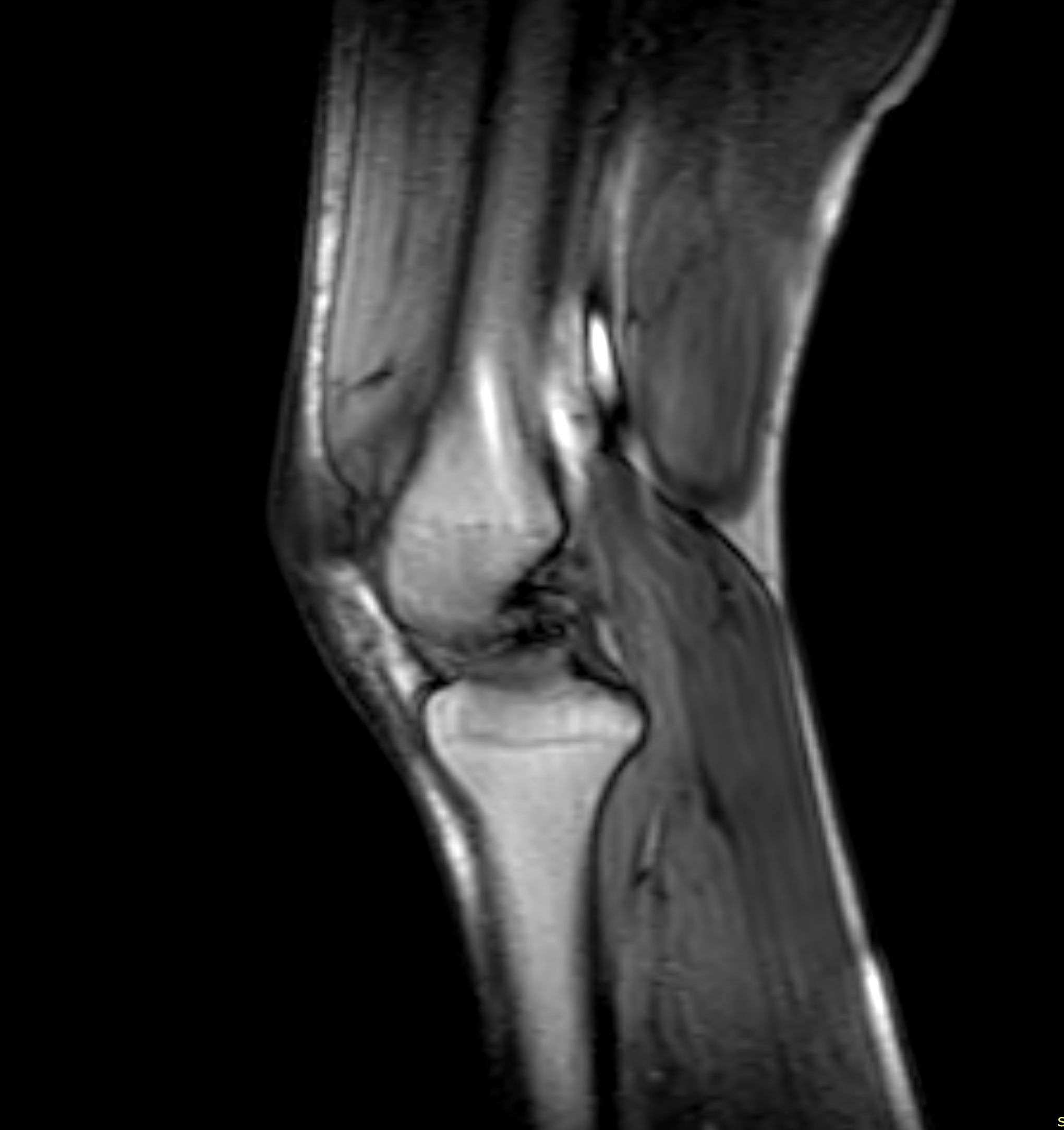
MRI knee for knee pain and suspected sports injury

MRI knee or X-ray – what is the difference?

MRI and standard X-ray show different structures and are therefore useful for different clinical questions.

X-rays primarily show bone and may be important when a fracture is suspected or when assessing certain degenerative changes.

MRI provides much more detailed information about the menisci, cruciate ligaments, other ligaments, cartilage, bone marrow, and many other soft tissues within the knee joint.

Following acute knee trauma, an X-ray may therefore be required first, while MRI can be used later if a soft tissue injury needs more detailed assessment.

Is contrast needed for a knee MRI?

For common questions such as meniscal tears, cruciate ligament injuries, collateral ligament injuries, or cartilage damage, a knee MRI is usually performed without intravenous contrast.

Contrast may be required for certain more specific clinical questions. This is determined according to the reason for the examination and the MRI protocol being used.

What happens during an MRI of the knee?

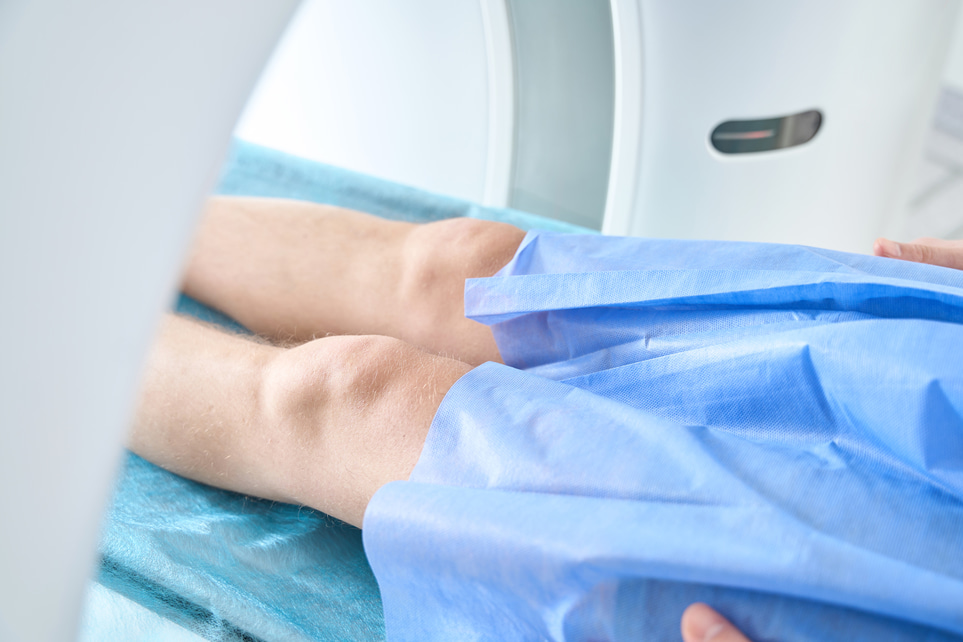
During the examination, you lie on your back with the knee positioned in a dedicated MRI coil that helps produce detailed images of the joint.

It is important to remain as still as possible while the images are being taken. The examination itself is painless, although the MRI scanner produces loud knocking and tapping sounds during parts of the scan. MRI does not use ionising radiation.

The total examination time depends on the MRI protocol being used. You will receive information about the examination and any necessary preparations beforehand.

When is a knee MRI not the best first examination?

MRI is not required for every type of knee pain. Short-term overuse symptoms, uncomplicated osteoarthritis, or certain acute injuries may need to be assessed in other ways first.

For suspected osteoarthritis, diagnosis is usually based mainly on symptoms and clinical examination, with standard X-rays used when appropriate. MRI is not routinely needed in uncomplicated cases.

Following a significant recent injury, an X-ray or urgent orthopaedic assessment may be more important than a planned MRI.

It is also important to remember that an MRI finding does not automatically mean that the abnormality is causing your symptoms or that surgery is necessary.

When should knee symptoms be assessed urgently?

Certain knee problems should be assessed promptly rather than through a routine planned MRI appointment.

This may apply if:

  • The knee is locked in one position and cannot be moved normally.
  • There is an obvious deformity after an injury.
  • The knee is severely unstable following trauma.
  • There is very marked swelling or suspected bleeding into the joint after injury.
  • There is concern about a fracture following significant trauma.

These situations may require prompt clinical assessment and, in some cases, different imaging or orthopaedic treatment.

Book an MRI of the knee

An MRI of the knee can provide detailed information about the menisci, cruciate ligaments, other ligaments, cartilage, bone marrow, and surrounding soft tissues. It can be particularly useful for persistent knee pain or when symptoms suggest an internal knee injury.

You do not need to decide on your own whether MRI is the most appropriate examination. A medical assessment is included as part of the booking process to review your symptoms and clinical question. If an X-ray, physiotherapy assessment, or another type of investigation is considered more appropriate, this can be recommended instead.

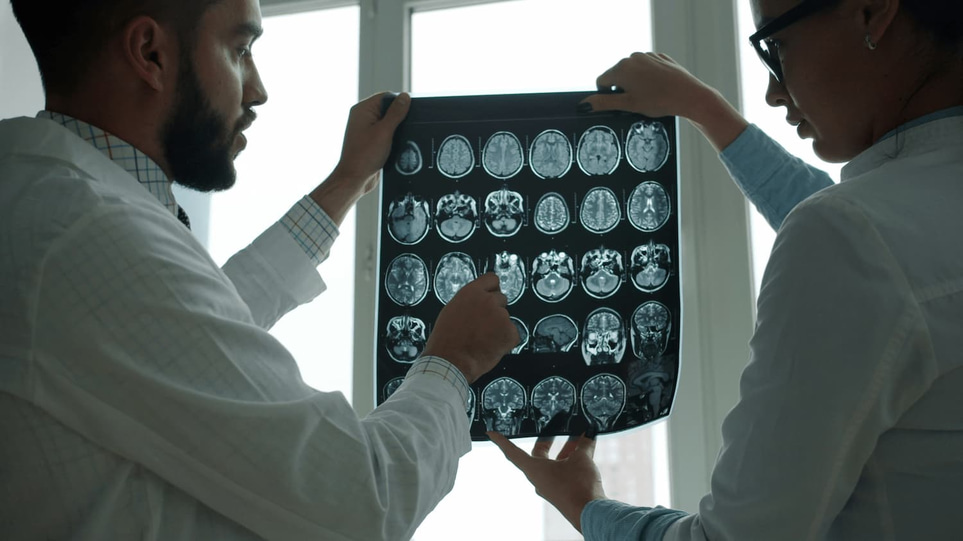
The MRI images are reviewed by a specialist radiologist, and the findings are summarised in a medical report.

The legs support the body's weight and make it possible to stand, walk, run and jump. The largest and strongest bone in the body is the femur, the knee joint is a complex structure that connects the thigh with the lower leg. The lower leg consists of the tibia and fibula, which lead down to the foot - a finely tuned construction that has many small bones and joints.

The foot is designed as an arch that absorbs shock and provides balance to the body. The Achilles tendon connects the calf muscles to the heel bone and is crucial for movement. Several important muscles, joints, ligaments and nerves work together to ensure that the legs function effectively both at rest and in activity. The nails on the toes protect against injury and pressure. Together, the legs constitute the body's most important locomotor apparatus.

KNE
Knee
The knee is the middle area on the front of the leg, between the thigh and lower leg, and covers both skin, muscles, tendons and the underlying kneecap.
Read more

After your order, we will send a referral for your MRI scan. Before that, you will have a brief consultation with a referring physician who will review your medical history and symptoms. After the consultation, your referral will be forwarded to the radiology clinic.

Magnetic Resonance Imaging (MRI) is a safe method for obtaining detailed images of the body's organs, tissues, and joints using a strong magnetic field and radio waves – completely without radiation.

Things to consider before the examination:

  • Clothing and items: You will be asked to remove clothing with metal, jewelry, watches, hearing aids, and other items that may be affected by the magnetic field.
  • Metal in the body: Inform the medical staff if you have a pacemaker, implants, metal clips, artificial joints, or other metal objects in your body.
  • Pregnancy: Notify the staff if you are pregnant or suspect that you might be.
  • Contrast agent: In some cases, a contrast agent is used to enhance image quality. You will be informed about any preparations or possible side effects if applicable.

After your MRI scan is completed, the images are reviewed by a specialist in radiology. The radiologist interprets the images based on the clinical question stated in your referral and prepares a medical report for the referring physician.

The referring physician from our team will then review the report and provide you with a written medical assessment and guidance.

If the examination reveals any abnormalities or findings, we will ensure that you are either referred to an appropriate healthcare provider or receive advice on suitable follow-up – all of this is included in the price.

Find the nearest test location

How does it work?

Order an MRI scan – four easy steps

Step 1

Order MRI

Select the MRI scan you wish to perform and add to your cart, for example lumbar spine, abdomen or full body. Complete your order and select a payment option, we always apply an open purchase on MRI for at least 30 days.

Step 2

Activate the referral

After payment, you’ll receive an email with instructions. Log in to enter your details and activate your referral. A physician will contact you to confirm your booking and discuss your preferences.

Step 3

Complete your MRI

You will be called via Kivra or mail within 7–10 working days, visit the clinic you have been called to at the appointed time. Contact the clinic directly for possible rescheduling if the time does not suit you. Read more

Step 4

Results and statement

You will receive feedback on your X-rays within 5–7 working days. However, in some cases it may take a few extra days before all results are ready and reviewed by specialist doctors. Read more

Your test answers in zample™

– A smarter tool for understanding your health

Select area

See the full list below

Other MRI examinations


MRI Full body Plus

MRI Full body Plus

MRI + blood tests

21 000 kr20 500 kr

MRI Full Body Pro

MRI Full Body Pro

MRI + blood tests + ECG

23 900 kr

Prostate Check – MRI & Stockholm3

Prostate Check – MRI & Stockholm3

MRI Prostate + Stockholm3

10 599 kr


MRCP

MRCP

MRI of the biliary tract and pancreas

7 695 kr

MRI Abdomen/MRCP

MRI Abdomen/MRCP

MRI of the abdomen

6 995 kr6 695 kr

MRI Aorta

MRI Aorta

MRI

9 495 kr

MRI Brain

MRI Brain

Magnetic resonance imaging

6 795 kr6 500 kr

5 299 kr

9 195 kr

9 195 kr

MRI Ears

MRI Ears

Magnetic resonance imaging

5 595 kr

13 995 kr

MRI Full Body

MRI Full Body

Magnetic resonance imaging

19 995 kr

MRI Full body Plus

MRI Full body Plus

MRI + blood tests

21 000 kr20 500 kr

MRI Full Body Pro

MRI Full Body Pro

MRI + blood tests + ECG

23 900 kr

MRI Kidneys

MRI Kidneys

MRI scan of the kidneys

6 195 kr

MRI Pancreas

MRI Pancreas

Magnetic resonance imaging

6 195 kr

MRI Pelvis

MRI Pelvis

MRI scan

6 495 kr

MRI Pituitary gland

MRI Pituitary gland

Magnetic resonance imaging

4 495 kr

6 395 kr

MRI Prostate

MRI Prostate

Magnetic resonance imaging

7 295 kr

7 295 kr

MR Auditory pathway

MR Auditory pathway

MRI of the pore

4 995 kr

MR Liver

MR Liver

MRI examination of the liver

6 795 kr


MRI Ankle

MRI Ankle

Magnetic resonance imaging

4 495 kr

MRI Cervical spine

MRI Cervical spine

Magnetic resonance imaging

5 995 kr

MRI Chest

MRI Chest

Magnetic resonance imaging

6 795 kr

MRI Elbow

MRI Elbow

Magnetic resonance imaging

4 495 kr

MRI face

MRI face

Magnetic resonance imaging

5 795 kr

MRI Femur

MRI Femur

Magnetic resonance imaging

4 495 kr

MRI Foot

MRI Foot

Magnetic Resonance Imaging

5 195 kr

MRI Forearm

MRI Forearm

Magnetic Resonance Imaging

3 795 kr

13 995 kr

MRI Full Body

MRI Full Body

Magnetic resonance imaging

19 995 kr

MRI Full body Plus

MRI Full body Plus

MRI + blood tests

21 000 kr20 500 kr

MRI Full Body Pro

MRI Full Body Pro

MRI + blood tests + ECG

23 900 kr

MRI Hand

MRI Hand

Magnetic resonance imaging

4 195 kr

MRI Hip joint and pelvis

MRI Hip joint and pelvis

Magnetic resonance imaging

5 795 kr

MRI Knee

MRI Knee

Magnetic Resonance Imaging

5 395 kr

MRI Knee

MRI Knee

Magnetic Resonance Imaging

4 895 kr

MRI Lower Leg

MRI Lower Leg

Magnetic resonance imaging

4 495 kr

MRI Lumbar spine

MRI Lumbar spine

MRI of the lumbar spine

5 995 kr

MRI Orbita

MRI Orbita

Magnetic resonance imaging

5 395 kr

MRI Sacroiliac joints

MRI Sacroiliac joints

Magnetic Resonance Imaging

4 495 kr

MRI Shoulder

MRI Shoulder

Magnetic resonance imaging

4 495 kr

MRI Sinus

MRI Sinus

Magnetic Resonance Imaging

6 495 kr

MRI Skull base

MRI Skull base

Magnetic Resonance Imaging

4 495 kr

MRI Temporomandibular joints

MRI Temporomandibular joints

Magnetic resonance imaging

5 495 kr

MRI Thoracic spine

MRI Thoracic spine

Magnetic resonance imaging

4 995 kr

MRI throat

MRI throat

Magnetic resonance imaging of the throat

6 195 kr

MRI Upper arm

MRI Upper arm

Magnetic Resonance Imaging

4 495 kr

MRI Wrist

MRI Wrist

Magnetic resonance imaging

4 495 kr

MR Achilles tendon

MR Achilles tendon

Magnetic resonance imaging

4 195 kr

5 595 kr

With us you are always safe with our satisfied customer guarantee!

With us you are always safe with our satisfied customer guarantee!

We want to help people improve their health. That's why it's important that you feel safe with us. As a healthcare provider, safety is our top priority and we always strive to deliver the highest quality in our services. If for any reason you are not completely satisfied with your experience with us - do not hesitate to contact us. We listen and will do what we can to make you feel safe and satisfied. Our satisfied customer guarantee is an expression of our commitment to you and your health - where we want to take our responsibility when you use our services to access your health data and improve your health.