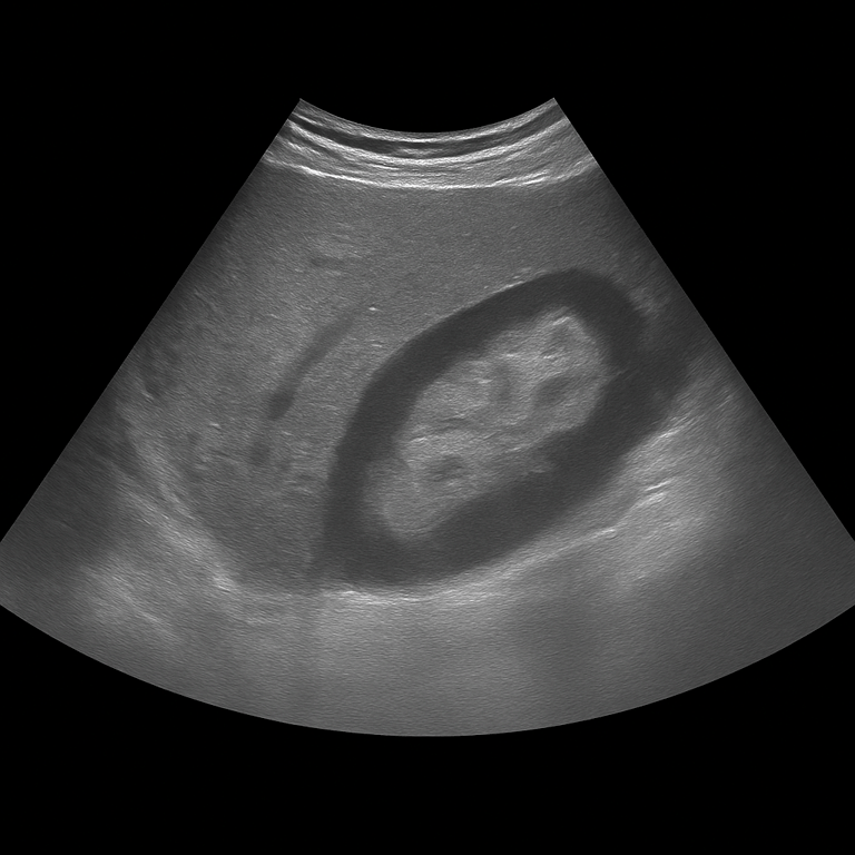
An ultrasound of the kidneys is used to assess the kidneys’ size, shape, tissue structure, and blood flow. The examination provides detailed real-time images that can reveal changes affecting kidney function or urinary drainage. It can be used to investigate conditions such as cysts, fluid accumulation, infections, or suspected tumors.
Ultrasound of the kidneys - for pain, urinary tract problems or altered blood tests
A kidney ultrasound is recommended for symptoms such as recurrent urinary tract infections, other urinary tract problems, or swelling in the body. The examination is also commonly performed when blood tests show abnormal kidney function markers (for example, elevated creatinine or cystatin C levels) to evaluate the appearance and drainage of the kidneys.
Unlike MRI and CT scans, which are often used for more advanced imaging or when tumors are suspected, ultrasound is the first-line method for assessing kidney structure and function. It can be performed without contrast agents and provides real-time visualization of the kidneys, making it possible to detect cysts or fluid accumulation without exposure to ionizing radiation.
Common symptoms and clinical indications
Recurrent urinary tract infections.
Elevated kidney function markers in blood tests.
Swelling of the legs or around the eyes (signs of fluid retention).
Follow-up of previous findings in the kidneys or urinary tract.
Conditions that can be detected with a kidney ultrasound
Fluid accumulation in the kidney (hydronephrosis) caused by urinary tract obstruction.
Kidney cysts or multiple cystic formations.
Changes in the kidney parenchyma associated with chronic kidney disease.
Kidney infection (pyelonephritis) causing swelling of the kidney tissue.
A tumor or suspicious mass in the kidney that may require further evaluation with MRI or CT.
Can kidney stones be seen on ultrasound?
In some cases, kidney stones located in the renal pelvis or the upper part of the ureter can be visualized on ultrasound. Kidney stone attacks occur when a stone becomes lodged in the ureter and blocks the flow of urine from the kidney. This creates increased pressure within the kidney and causes severe pain, typically felt in the flank and sometimes radiating toward the abdomen or groin.
If kidney stones are suspected, computed tomography (CT) is the radiological first-line imaging method, and ultrasound is rarely used as the primary diagnostic tool. If you suspect that you are experiencing an ongoing kidney stone attack, this examination is not recommended. Instead, you should seek immediate medical attention at your nearest emergency department.
How an ultrasound of the kidneys is performed
The examination is performed while you lie on your back or side. A gel is applied to the skin and the doctor moves the ultrasound probe over the area where the kidneys are located. Both kidneys are assessed in longitudinal and cross-sectional views, and if necessary, the bladder is also examined to assess any impact on urine flow. The examination is painless and usually takes 15–20 minutes. You do not normally need to fast or prepare, but sometimes the doctor may ask you to drink water before the examination so that the bladder is slightly full.
Order an ultrasound examination of the kidneys - get a report and recommendation from a doctor
The images are reviewed by a specialist in radiology who draws up a written medical report. The answer is delivered digitally within a few working days and can be shared with your treating doctor for further investigation or treatment. If necessary, the findings can be followed up with MRI or CT for further mapping.