Quick version
Ultrasound uses high-frequency sound waves without radiation to create real-time images of the body's organs, blood vessels, and soft tissues. The examination usually takes 15 to 35 minutes and is performed by applying a gel to the skin before moving a transducer over the area. The method is completely safe and highly valuable for conditions such as pregnancy, abdominal pain, lumps, or suspected blood clots. Because sound waves are obstructed by bone and intestinal gas, ultrasound is often combined with blood tests or other imaging techniques to provide a complete picture.
Ultrasound is an examination that many people encounter at some point in their lives – for abdominal pain, a lump in the throat, swollen legs or during pregnancy. For many, it raises the same question: what do you actually see, and how does an ultrasound examination work in practice? The answer is often reassuring. Ultrasound is a fast, gentle and well-proven method that can provide important information directly while the examination is in progress.
What is ultrasound and what is it used for?
Ultrasound is a medical imaging method that uses high-frequency sound waves to create images of the body's internal organs and tissues. A small handheld transducer, often called a probe, sends out sound waves and receives echoes that are converted into images on a screen. Unlike X-rays and computed tomography, ultrasound does not use ionizing radiation.
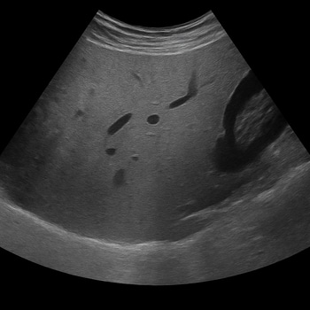
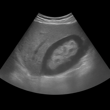
The method is used in many different situations. Common examples are examinations of liver, gallbladder, kidney, thyroid, blood vessels, soft tissues and pelvic organs. Ultrasound is also used to assess blood flow with a so-called Doppler examination and to guide needles when, for example, taking samples or draining.
For the patient, the benefit is often tangible. In the case of pain under the right rib cage, ultrasound can show gallstones. In the case of a lump on the neck, it can be assessed whether the change is in the thyroid gland, a lymph node or in the soft tissues. In the case of swelling in a leg, the examination can help the doctor detect or rule out a blood clot in the deep veins. Such answers often directly affect the next step.
How is an ultrasound examination performed?
How an ultrasound examination is performed depends on which part of the body is being examined. For an ultrasound outside the body, you will usually be asked to remove clothing and jewelry over the area to be assessed and lie down on a table. The examiner will then apply a gel to your skin to ensure good contact between the probe and your body.
The gel may feel cold, but the examination itself is usually not painful. The probe is slowly moved over the skin while images are displayed in real time on the screen. In some situations, the examiner may need to press a little harder to see an organ clearly, which can feel uncomfortable if the area is already tender.
Sometimes ultrasounds are performed through the body's openings, such as vaginally or through the rectum. In this case, a narrower probe is used and the examination often provides better images of the pelvic organs or prostate than if only the outside of the body is examined. Before some examinations, you may need to fast for a few hours or have a full bladder, as this improves the ability to see the right structures.
The examination itself usually takes between 15 and 35 minutes, depending on what is to be examined. Since your images first need to be assessed and reviewed by a responsible radiologist, you will not receive a final result immediately. As soon as the assessment is complete, your answer will be compiled and returned to you via us. If your results require further medical follow-up, we will help you with recommendations for the next step.
Ultrasound during pregnancy, abdominal discomfort and other common questions
Many people associate ultrasound with pregnancy, and it is also the most common situation in which healthy people encounter the method. Ultrasound is the preferred imaging method for monitoring pregnancy because it can show fetal development without using radiation. It is used, among other things, to assess the length of pregnancy, the position of the placenta and the growth of the fetus.
But ultrasound is at least as important outside of pregnancy care. In the case of abdominal pain, the examination can provide guidance about the bile ducts, liver, kidneys and sometimes the bladder or large blood vessels. If kidney stones are suspected, ultrasound can sometimes show whether urine flow is obstructed, although other methods are sometimes needed for the full picture.
Another common question is whether ultrasound can be used for the heart and blood vessels. The answer is yes. Ultrasound can assess blood flow in arteries and veins, and with Doppler technology you can see how the blood moves and sometimes also hear the flow. This makes the method useful for, for example, atherosclerosis in the carotid arteries, venous insufficiency or suspected deep vein thrombosis.
Is ultrasound dangerous? Benefits, limitations and safety
For diagnostic use, ultrasound has a strong safety profile. Standardized medical ultrasound has no known harmful effects on humans when used correctly in healthcare, and since the method is not based on ionizing radiation, the radiation exposure that is present in some other imaging examinations is avoided.
This does not mean that ultrasound is always the best method. The sound waves are disturbed by air and gas, which makes the intestines and air-filled lungs often difficult to assess. Ultrasound also does not pass through bones in the same way as through soft tissue, so if the doctor needs to see inside bones or certain joints, a magnetic resonance imaging or other imaging diagnostics may be more appropriate.
Body structure can also affect image quality. If structures are deep or if a lot of tissue dampens the sound waves, the images are sometimes less clear. A normal ultrasound therefore does not always rule out disease completely, but the answer must be interpreted together with symptoms, examination findings and sometimes blood tests. This is an important patient question: a "no abnormalities" answer is valuable, but not always the end point of the investigation.
When is ultrasound enough - and when is blood tests or other testing needed?
Ultrasound shows structure and movement, but it does not tell everything about how the body functions at the cellular level or whether metabolism is disturbed. For example, a liver can look normal on ultrasound even though liver function tests in the blood is elevated. Similarly, thyroid gland can have a normal appearance even though hormone levels show over- or under-function. That is why ultrasound is often combined with blood tests to provide a more reliable overall picture.
If you have been told that an ultrasound is normal but you still feel unwell, it is reasonable to ask what the next step is. In some cases, this involves new tests, in others, follow-up, other imaging diagnostics or assessment by a specialist.
Ultrasound is therefore not just a technique for “looking inside the body”, but a way to make better medical decisions without unnecessary burden on the patient. This makes the examination particularly valuable when you need quick answers, follow a course over time or determine whether more advanced investigation is really needed. Used correctly, it can reduce both anxiety and waiting.