Quick version
Aortic aneurysm is a weakening and widening of the body's main artery, which usually develops without symptoms for a long time. The condition is often discovered by chance and requires regular follow-up to reduce the risk of complications. Most aneurysms are small and harmless, but the risk of rupture increases as the diameter increases.
When a rupture occurs, an acute and life-threatening situation occurs with severe pain and internal bleeding. Early detection, control of risk factors and correct treatment are crucial for the prognosis. Lifestyle changes and medical treatment play an important role in reducing the risk of progression and complications.
What is an aortic aneurysm?
An aortic aneurysm is a dilation of the body's largest artery, the aorta. The condition is also called an aortic aneurysm and occurs when the vessel wall weakens and gradually stretches. The aorta transports oxygen-rich blood from the heart to the rest of the body and is located deep in the chest and abdomen, which means that changes often occur without giving clear symptoms.
Most aortic aneurysms develop slowly over many years and are often discovered by chance during examinations for other reasons. In many cases, the condition is completely asymptomatic until it becomes large or ruptures.
How is the aorta structured?
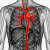
The aorta can be compared to a strong artery that originates from the left ventricle of the heart. It is anatomically divided into several parts: the ascending aorta (the ascending part), the aortic arch (arcus), the descending aorta (the descending part), as well as the thoracic aorta in the chest and the abdominal aorta in the abdomen. Aneurysm can occur in all of these parts, but is most common in the abdominal aorta.
Why does an aortic aneurysm occur?
The underlying cause is a weakening of the connective tissue in the vessel wall. When the vessel wall loses its elasticity, it becomes less resistant to the pressure created by blood flow. This leads to the vessel gradually widening.
Several factors can contribute to this weakening:
- Aging and natural breakdown of the vessel wall
- High blood pressure that puts strain on the vessels over time
- Smoking, which is one of the strongest risk factors
- Arteriosclerosis
- Heredity and genetic connective tissue diseases
Different types of aortic aneurysm
Aortic aneurysms are divided depending on where in the body they are located:
- Abdominal aortic aneurysm (AAA): The most common form, located in the abdomen.
- Thoracic aneurysms: Occur in the chest and are less common.
- Ascending aortic aneurysm: Located near the heart and may be linked to hereditary conditions.
Thoracic aneurysms are more difficult to detect with ultrasound and are more often diagnosed with computed tomography (CT) or magnetic resonance imaging (MRI).
Symptoms often consist of a silent condition
Most aortic aneurysms do not cause any symptoms at all. Because the aorta is protected deep within the body, a dilation is rarely noticed until it has become large.
In some cases, larger aneurysms can cause diffuse symptoms, such as:
- A feeling of pressure or pain in the stomach or back
- A pulsating feeling in the abdomen
- Discomfort in the chest (in the case of thoracic aneurysms)
Symptoms of aortic rupture, an emergency condition
If an aortic aneurysm ruptures, a life-threatening situation arises that requires immediate care.
Common symptoms of abdominal rupture are:
- Sudden and very severe abdominal pain
- Back pain that may radiate to the groin
- A distended abdomen
- Nausea and vomiting
- Dizziness, fainting or shock
If a rupture occurs in the chest, the symptoms may resemble a heart attack, with intense pain in the chest and back.
If a rupture is suspected, you should always call 112.
How to detect an aortic aneurysm early
Aortic aneurysms are usually discovered by chance during examinations performed by other causes, as the condition rarely causes symptoms in the early stages. Imaging methods are used to confirm the diagnosis and assess the size and spread of the aneurysm.
Ultrasound is the most common method for detecting and monitoring abdominal aortic aneurysms. The examination is quick, gentle and is also used in screening programs. It provides a good estimate of the diameter of the vessel, which is crucial for further management.
For a more detailed assessment, computed tomography of the aorta CT aorta, is used, which is the most important method when a larger aneurysm is suspected or before treatment. The CT examination provides a very accurate image of the entire aorta and shows the aneurysm's exact size, shape and relationship to other blood vessels. It is also used in acute conditions, for example if it is suspected that the aneurysm has ruptured.
With CT aorta, contrast agents are usually given into the blood, which makes the vessels appear clearly. This enables precise mapping that is necessary before planning surgery or other treatment.
Magnetic resonance imaging (MRI) may be used as an alternative in some cases, especially when radiation is avoided or additional information is needed. For thoracic aneurysms, CT or MRI are often necessary, as these cannot be adequately assessed with ultrasound. The choice of examination method is based on the location of the aneurysm, its size, and whether the situation is urgent or planned.
When does an aneurysm need to be treated?
An abdominal aortic aneurysm is usually defined as a diameter of 30 mm or more. Most aneurysms are small and pose a low risk of rupture. Therefore, they are not treated immediately but are monitored regularly with ultrasound.
As the aneurysm grows, the risk of rupture increases. At a diameter of approximately 55 mm in men (often slightly smaller in women), surgery is considered, where the weakened part of the vessel is replaced or reinforced.
Decisions on treatment are always based on a comprehensive assessment of risks, symptoms and the patient's general health.
Follow-up and checks
People with smaller aneurysms are followed regularly in healthcare, usually via vascular surgery units. The checks aim to monitor growth and determine when treatment may be appropriate.
Many patients are also offered preventive treatment with drugs, such as blood pressure-lowering, lipid-lowering and platelet-inhibiting drugs to reduce the risk of cardiovascular disease.
Risks and prognosis
Small aortic aneurysms generally have a low risk of rupture and can often be followed for a long time without intervention. However, the risk increases as the aneurysm grows.
A rupture is a life-threatening condition with a high mortality rate, but the prognosis improves significantly if diagnosed and treated in time. With modern methods, more and more patients can be saved.
Living with an aortic aneurysm
Being diagnosed with an aortic aneurysm can be worrying, but many people live long lives without it causing problems. Regular check-ups and medical follow-up provide good security.
It is also important to reduce the strain on the vessels through lifestyle changes:
- Quit smoking
- Treat high blood pressure
- Control blood lipids
- Be physically active
When contacting healthcare, it is important to always inform that you have or have had an aortic aneurysm.
Heredity and screening
There is a hereditary component in some cases. If you have a close relative with an aortic aneurysm, the risk may be increased, and it may be appropriate to discuss an examination with a doctor.
In Sweden, screening for abdominal aortic aneurysm is offered to men in certain age groups, since early detection can prevent serious complications.